Diagnostic Musculoskeletal Ultrasound
Cutting-Edge MSK Imaging—Right in Our Office.
At Sierra Spine & Fitness, treatment starts with diagnostic certainty. Before recommending care, we want to understand which tissues are involved, how significant the injury may be, and what factors are contributing to your symptoms.
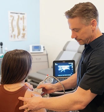
Performing ultrasound with a patient
Diagnostic musculoskeletal ultrasound is a cutting-edge imaging technology that allows us to examine many muscles, tendons, ligaments, joints, bursae, and peripheral nerves in real time. We use ultrasound in conjunction with a detailed clinical examination and, when necessary, digital X-rays or referral for additional imaging. By combining these findings, we can develop a more precise diagnosis and a treatment plan tailored to your condition.
What Is Musculoskeletal Ultrasound?
Musculoskeletal ultrasound uses high-frequency sound waves to create detailed images of structures beneath the skin. Unlike an X-ray, which is primarily used to evaluate bones and joint alignment, ultrasound is especially useful for examining many of the soft tissues that commonly produce pain.
Ultrasound can identify:
- Tendinopathy and tendon degeneration
- Partial or complete tendon tears
- Muscle strains and tears
- Ligament injuries
- Bursitis and joint swelling
- Calcium deposits within tendons
- Ganglion cysts and other soft-tissue masses
- Peripheral nerve enlargement or entrapment
- Tissue thickening, scarring, or inflammation
- Changes associated with conditions such as plantar fasciitis and carpal tunnel syndrome
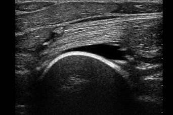
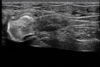
Real pathology, on the screen.
These are examples of the kind of findings diagnostic ultrasound can reveal—information that helps guide a more focused, individualized treatment plan.


The Advantage of Real-Time, Dynamic Imaging
One of the greatest advantages of diagnostic ultrasound is its ability to evaluate tissues while they move. Instead of viewing only a still image, we can observe a tendon gliding, a muscle contracting, or a nerve moving through its surrounding tissues.
This dynamic assessment may help reveal problems that are difficult to appreciate when the body is completely still. We can also compare the painful area with the unaffected side during the same examination.
Immediate Information Without Radiation
Diagnostic ultrasound does not use ionizing radiation. It is noninvasive, generally comfortable, and can often be performed during the same visit as your clinical examination.
Because the images are produced in real time, Dr. Roberts, DC can correlate what appears on the screen with your exact area of pain and with the movements that reproduce your symptoms. When appropriate, the images can also be used to document the condition and monitor changes over time.
Ultrasound, Examination, and Digital X-Ray
No single test tells the entire story. Imaging findings must be interpreted in the context of your symptoms, history, strength, mobility, neurological function, and orthopedic examination.
Ultrasound provides detailed information about many soft tissues, while digital X-ray is better suited to evaluating bones, arthritis, alignment, and certain types of trauma. Using the appropriate tools together helps us determine not only what appears abnormal, but whether that finding is likely to explain your symptoms.
At Sierra Spine & Fitness LLC, our diagnostic process may combine:
- A detailed history and physical examination
- Orthopedic and neurological testing
- Functional movement assessment
- Diagnostic musculoskeletal ultrasound
- Digital X-ray, when clinically indicated
- Referral for MRI or another specialized study when necessary
Areas We Commonly Evaluate
Diagnostic musculoskeletal ultrasound can be used to examine many regions of the body. The usefulness of ultrasound depends on the body region and the suspected condition—deeper structures and tissues located behind bone may require another type of imaging.
Shoulder
Rotator cuff tendons, biceps tendon, bursae, and calcific deposits.
Elbow
Common extensor and flexor tendons, distal biceps, distal triceps, and ulnar nerve.
Wrist & hand
Carpal tunnel, tendons, ligaments, cysts, and small joints.
Hip
Superficial tendons, bursae, and surrounding soft tissues.
Knee
Quadriceps and patellar tendons, collateral ligaments, joint fluid, and selected superficial structures.
Ankle & foot
Achilles tendon, plantar fascia, ankle tendons, ligaments, and selected peripheral nerves.
Imaging That Helps Guide Treatment
A clearer understanding of the injured tissue allows treatment to be more focused. Ultrasound findings may help us determine whether your condition is most appropriate for rehabilitation, activity modification, focused or radial shockwave therapy, another non-surgical treatment strategy, or referral for additional medical evaluation.
When follow-up imaging is clinically useful, ultrasound may also allow us to observe changes in the tissue and adjust your treatment plan accordingly.
Common conditions diagnostic ultrasound helps clarify
Find out if diagnostic ultrasound is right for you.
If you have persistent pain, weakness, swelling, numbness, or an injury that has not responded as expected, diagnostic musculoskeletal ultrasound may provide valuable information. Contact Sierra Spine & Fitness LLC to schedule an evaluation.
582 Searls Ave, Nevada City, CA 95959
- Monday9:00am – 1:00pm, 2:00pm – 6:00pm
- Tuesday9:00am – 1:30pm, 2:00pm – 4:00pm
- Wednesday9:00am – 1:00pm, 2:00pm – 6:00pm
- ThursdayClosed
- Friday9:00am – 4:00pm
- Saturday & SundayClosed